Condition Focus: Rheumatoid Arthritis — Synoviocyte-Specific Inflammation
Most PBM studies in joint disease measure broad outcomes — overall pain scores, general inflammatory markers, tissue histology. This study from Ryu and colleagues goes deeper, examining PBM’s effects specifically on fibroblast-like synoviocytes (FLS) — the specialised cells that line the inner surface of the joint capsule and play a central role in joint inflammation.
Using both cell culture (in vitro) and animal RA models (in vivo), the researchers demonstrated that PBM directly reduces inflammatory parameters in FLS cells. This is significant because FLS cells are not passive bystanders in joint disease — they actively produce inflammatory mediators, recruit immune cells, and drive the tissue destruction that leads to permanent joint damage.
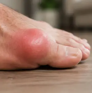
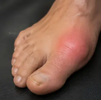
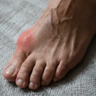
For gout, the synovium is ground zero. Monosodium urate crystals preferentially deposit in the synovial fluid and synovial membrane, triggering the inflammatory cascade directly in the tissue that FLS cells populate. The fact that PBM can modulate FLS cell behaviour — reducing their inflammatory output without destroying them — means it has the potential to address gout inflammation at the exact tissue layer where the disease process begins.
This study also bridges the gap between anti-inflammatory evidence (which shows PBM reduces broad inflammatory markers) and tissue-specific evidence (which shows exactly where in the joint that reduction occurs). For a device designed to be applied externally over a joint, knowing that the light’s effects reach and modulate the synovial lining is critical validation.
G.O.A.T. for Gout Alignment:
The G.O.A.T.’s 850 nm wavelength is selected specifically for its tissue penetration depth — sufficient to reach the synovial membrane of the MTP joint through overlying skin, subcutaneous tissue, and joint capsule. This study confirms that PBM modulates the exact cell type that populates the tissue where urate crystals deposit.
Link to original research here
Editor’s note: The synoviocyte-specific evidence here complements the broader neutrophil and inflammatory mediator data in Alves et al 2014. For the complete four-cytokine panel (IL-1β, IL-6, TNF-α, PGE₂) produced by synovial tissue in joint inflammation, see Tomazoni et al 2017. The COX-2/PGE₂ pathway through which synoviocytes drive pain is explored in Albertini et al 2007. For the cytokine profile modulation at tissue level in OA, see PBM on OA Narrative Review 2025.
Related Articles
- Effect of PBM on Inflammatory Mediators and Neutrophils in Joint Inflammation – Alves et al 2014
- Effects of PBM on Inflammatory Response in Experimental OA – Tomazoni et al 2017
- Anti-Inflammatory Effect of PBM on COX-2 and PGE₂ in Joint Inflammation – Albertini et al 2007
- Effects of PBM on OA from In Vivo and In Vitro Studies – Narrative Review 2025
- PBM Mitigates Chondrocyte Catabolism via Macrophage M1 Polarization – 2025
Key Takeaways
- PBM directly modulates inflammation in fibroblast-like synoviocytes — the cells lining the joint cavity
- Synovium is the exact tissue where urate crystals deposit and trigger gout flares
- Both in vitro (cell culture) and in vivo (animal model) evidence provided
- Demonstrates PBM reaches and modulates the tissue layer most relevant to gout pathology
Study Overview
| Study Type: | Animal study + in vitro (cell culture) |
| Wavelength(s): | Not specified in abstract |
| Treatment Protocol: | FLS cell culture + RA animal model treatment |
| Sample Size: | Cell culture + animal groups |
| Primary Outcome: | Reduced inflammatory parameters in fibroblast-like synoviocytes |
Full Citation
Ryu JH, et al. (2023). Photobiomodulation ameliorates inflammatory parameters in fibroblast-like synoviocytes and experimental rheumatoid arthritis models. International Journal of Molecular Sciences. View Publication