Condition Focus: Cartilage Matrix Synthesis — Human Chondrocyte Evidence
Most cartilage repair studies use animal cells or animal models. This 2025 study from Oliveira and colleagues, published in Nature’s Scientific Reports, used primary human chondrocytes — the actual cells responsible for producing and maintaining cartilage in human joints. This distinction matters because animal chondrocytes can behave differently from human cells, making human cell data more directly relevant to clinical device applications.
The researchers tested LED wavelengths from 600 to 940 nm and found that 850 nm and 940 nm near-infrared produced the strongest results. Collagen II — the primary structural protein of articular cartilage — increased by 1.3 to 1.5 times over untreated controls. Aggrecan — the proteoglycan responsible for cartilage’s compressive resilience and water retention — increased by 1.5 to 2.1 times. Both increases were achieved without compromising cell viability, and daily or every-other-day treatment schedules were effective.
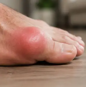
For gout, cartilage damage is a progressive consequence of repeated flares. Each flare deposits urate crystals in the synovial space, triggering inflammatory cascades that degrade cartilage through MMP activity and inflammatory cytokine-driven catabolism. Over years, this leads to the joint erosion and deformity seen in chronic tophaceous gout. Evidence that PBM can stimulate the very matrix molecules that cartilage needs to rebuild — using human cells at wavelengths matching the G.O.A.T. — supports the device’s role in long-term joint protection.
G.O.A.T. for Gout Alignment:
The G.O.A.T.’s 850 nm wavelength matches the optimal NIR wavelength identified in this study. The Collagen II and Aggrecan upregulation demonstrated here directly supports the G.O.A.T.’s potential for chronic gout management — not just acute flare relief but long-term cartilage maintenance between flares.
Link to original research here
Editor’s note: This human chondrocyte evidence complements the cartilage integrity restoration demonstrated in the animal OA model by Balbinot et al 2021. The macrophage-chondrocyte cross-talk that protects cartilage through M1→M2 shift is mapped in PBM and Chondrocyte Catabolism 2025. The MMP-13 suppression that reduces cartilage degradation is confirmed in the meta-analysis by Nambi 2021. For the comprehensive parameter tables for cartilage regeneration, see PBM in Cartilage Regeneration 2025.
Related Articles
- PBM Restores Cartilage Integrity and Reduces Chronic Pain in OA – Balbinot et al 2021
- PBM Mitigates Chondrocyte Catabolism via Macrophage M1 Polarization – 2025
- LLLT Effects on Inflammatory Biomarkers in OA: Meta-Analysis – Nambi 2021
- PBM in Promoting Cartilage Regeneration – 2025
- PBM, Cells of Connective Tissue and Repair – Houreld et al 2022
Key Takeaways
- Human chondrocytes — not animal cells — used for direct clinical relevance
- 850 nm and 940 nm NIR produced optimal results: ColII ↑1.3-1.5x, Aggrecan ↑1.5-2.1x
- Cell viability maintained — PBM stimulates repair without harming cells
- Published in Nature Scientific Reports — high-visibility journal
Study Overview
| Study Type: | In vitro (primary human chondrocytes) |
| Wavelength(s): | 600–940 nm LED; 850 nm and 940 nm most effective |
| Treatment Protocol: | Daily or every other day, up to 4 min per session |
| Sample Size: | Primary human chondrocyte cultures |
| Primary Outcome: | Collagen II ↑1.3-1.5x; Aggrecan ↑1.5-2.1x at 850/940 nm |
Full Citation
Oliveira S, et al. (2025). Near-infrared photobiomodulation stimulates viability and cartilage matrix synthesis in human chondrocytes. Scientific Reports, 15, 28792. View Publication