Condition focus: Blue Light Toxicity & Retinal Cell Death Pathways
Blue light exposure from digital devices and LED lighting poses increasing concern for retinal health, yet the molecular pathways linking blue light to retinal cell death remain incompletely understood. This study investigated the role of p53, a master regulator of cellular stress responses and apoptosis, in mediating blue light-induced retinal damage. Using human retinal cell cultures and animal models, researchers examined p53 activation, downstream apoptotic signaling, and oxidative stress markers following blue light exposure, with comparison to protective red light wavelengths.
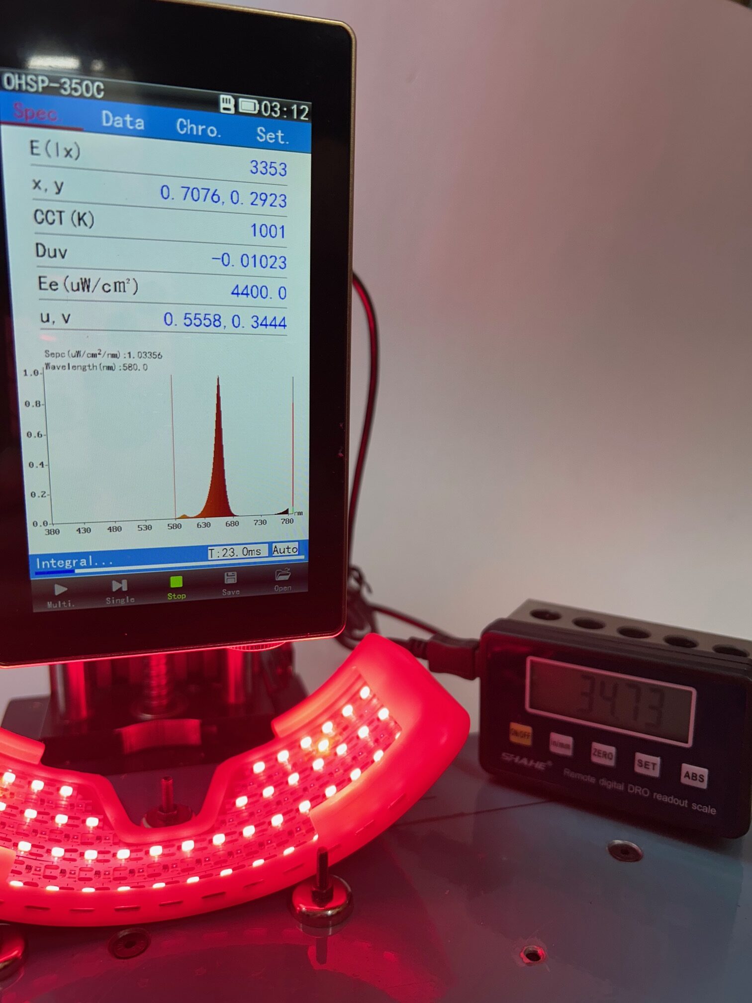
Results demonstrated that blue light exposure rapidly activated p53 through oxidative stress-induced DNA damage, triggering transcription of pro-apoptotic genes including BAX, PUMA, and NOXA. p53 activation correlated with mitochondrial dysfunction, cytochrome c release, and caspase activation leading to retinal cell death. Importantly, cells lacking functional p53 showed reduced sensitivity to blue light toxicity, confirming p53’s central role in the death pathway. In contrast, red light (630-670 nm) suppressed p53 activation by reducing oxidative stress and enhancing mitochondrial antioxidant defenses. Combined exposure to red and blue light significantly attenuated p53-mediated apoptosis compared to blue light alone. The findings establish p53 as a critical mediator of blue light retinal toxicity and validate red light photobiomodulation as a protective countermeasure through p53 pathway suppression.
WaveFront Alignment:
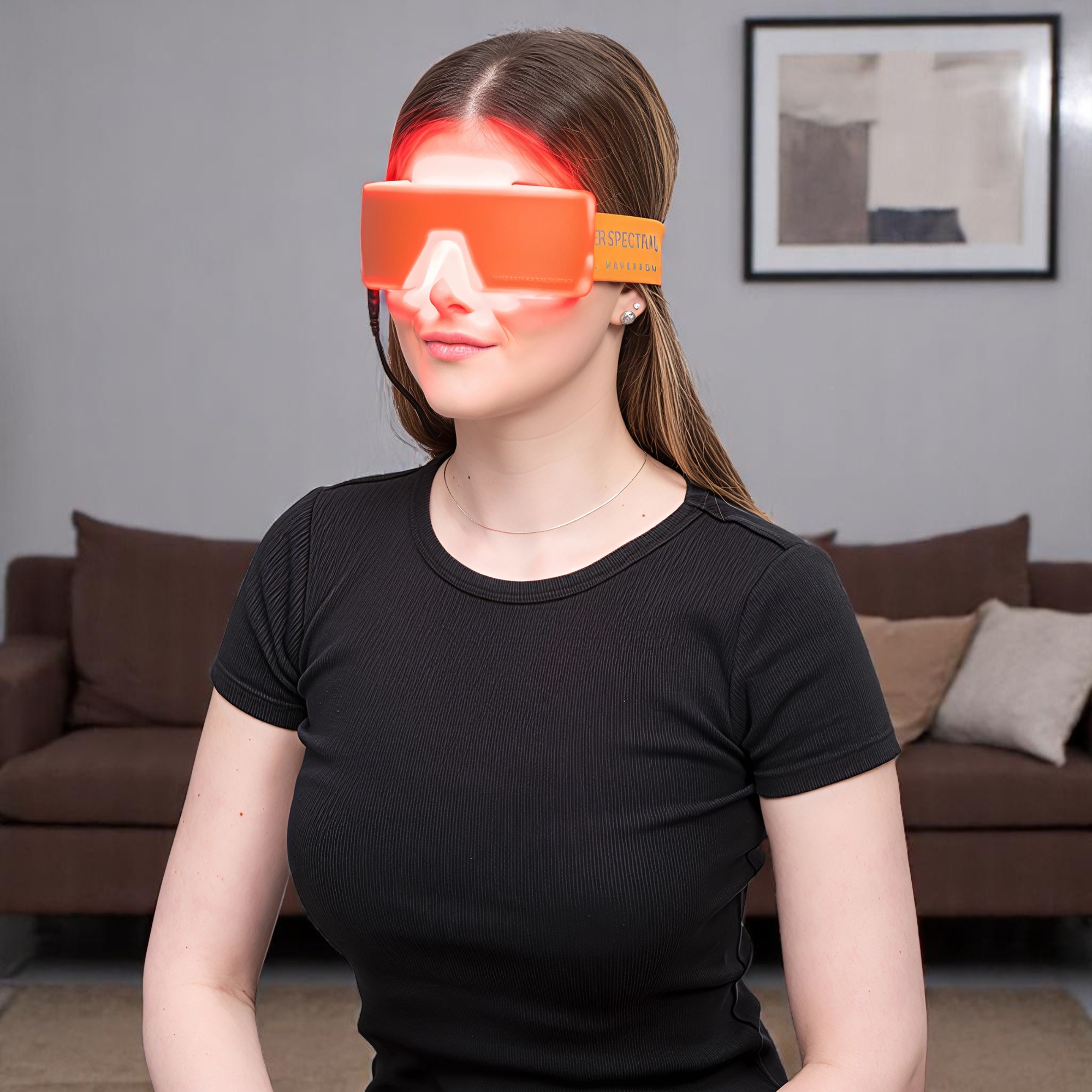
Fietz’s identification of p53-mediated apoptosis in blue light toxicity provides molecular-level validation for the Spectral WaveFront’s protective potential against modern environmental light stress through red light’s suppression of oxidative stress-induced cell death pathways.
Read full article here
Editor’s note: Fietz 2023 elucidates p53-mediated pathways in blue light retinal toxicity. For related blue light damage mechanisms, see Cheng 2021 and broader light effects in Osborne 2016. Red light protection against blue light demonstrated in Nunez-Alvarez 2019. Mitochondrial oxidative stress mechanisms in Fitzgerald 2010.
Related Articles
- Blue Light, Oxidative Stress, and Autophagy in RPE – Cheng 2021
- Visual Light Effects on Mitochondria – Osborne 2016
- Blue Light Exacerbates and Red Light Counteracts Negative Insults – Nunez-Alvarez & Osborne 2019
- NIR Reduces Oxidative Stress in Optic Nerve Injury – Fitzgerald 2010
- Mitochondrial Alterations of RPE in AMD – Feher 2006
Key Takeaways
- Blue light rapidly activated p53 through oxidative stress-induced DNA damage, triggering pro-apoptotic gene transcription
- p53 activation led to mitochondrial dysfunction, cytochrome c release, and caspase-mediated cell death
- Red light (630-670 nm) suppressed p53 activation by reducing oxidative stress and enhancing antioxidant defenses
- Combined red/blue light exposure significantly attenuated p53-mediated apoptosis versus blue light alone
Study Overview
| Study Type: | In vitro and in vivo mechanistic research |
| Wavelength(s): | Blue light (toxic) vs Red light (630-670 nm, protective) |
| Treatment Protocol: | Light exposure in retinal cell cultures and animal models with p53 pathway analysis |
| Sample Size: | Human retinal cell cultures and animal models |
| Primary Outcome: | p53 mediates blue light toxicity; red light suppresses p53 activation and protects cells |
Full Citation
Fietz AG. (2023). Blue light damage and p53: unravelling the role of p53 in oxidative stress-induced retinal apoptosis. Photochem Photobiol Sci, 22(4):857-869. View Publication